

Ultrasound does not use ionizing radiation, but it does introduce acoustic energy into the body. FDA and professional guidance therefore emphasize prudent use: trained operators, appropriate equipment settings, attention to the thermal and mechanical indices, and exposure time limited to the purpose of the scan. Elective imaging should remain clearly non-diagnostic and supplemental to regular prenatal care.
Why Safety Guidance Matters in Elective Ultrasound
3D, 4D, and HD ultrasound can give families a memorable view of their baby and create a welcoming opportunity for family connection. Responsible studios support that experience with thoughtful scanning practices and clear communication about what an elective session can and cannot provide.
Safety is not a slogan or a single machine setting. It is a working process that combines operator education, purposeful scanning, appropriate presets, equipment care, infection-control procedures, and respect for the relationship between elective imaging and prenatal healthcare.
This guide explains the main principles in plain language for both expecting parents and people preparing to operate an elective ultrasound studio.
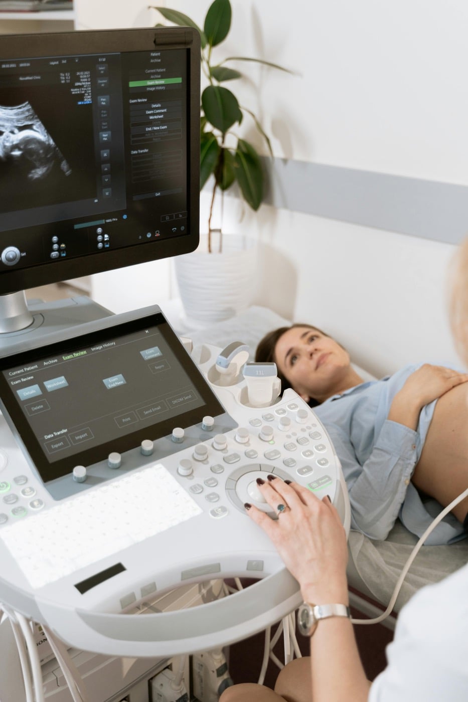
Three Facts That Create a Better Safety Conversation
Accurate wording helps families make informed decisions and helps studio teams build consistent operating procedures.
No Ionizing Radiation
Ultrasound forms images using high-frequency sound waves. Unlike X-ray and CT imaging, it does not expose a person to ionizing radiation.
Energy Still Requires Care
Ultrasound energy can produce thermal and mechanical effects. That is why operator training, displayed output indices, dwell time, and total scanning time matter.
Purpose Defines the Service
A diagnostic examination answers medical questions. An elective session is a non-diagnostic viewing and keepsake experience that supplements prenatal care.
The FDA describes ultrasound as having an excellent safety record and very low risks when used prudently by appropriately trained providers. Its guidance also notes the potential for biological effects, warns that unnecessary prolonged exposure or use by untrained operators may increase risk, and states that ultrasound solely for nonmedical keepsake purposes has been discouraged. A responsible studio should understand this published position, avoid overstating safety, and make conservative operation central to training and policy.
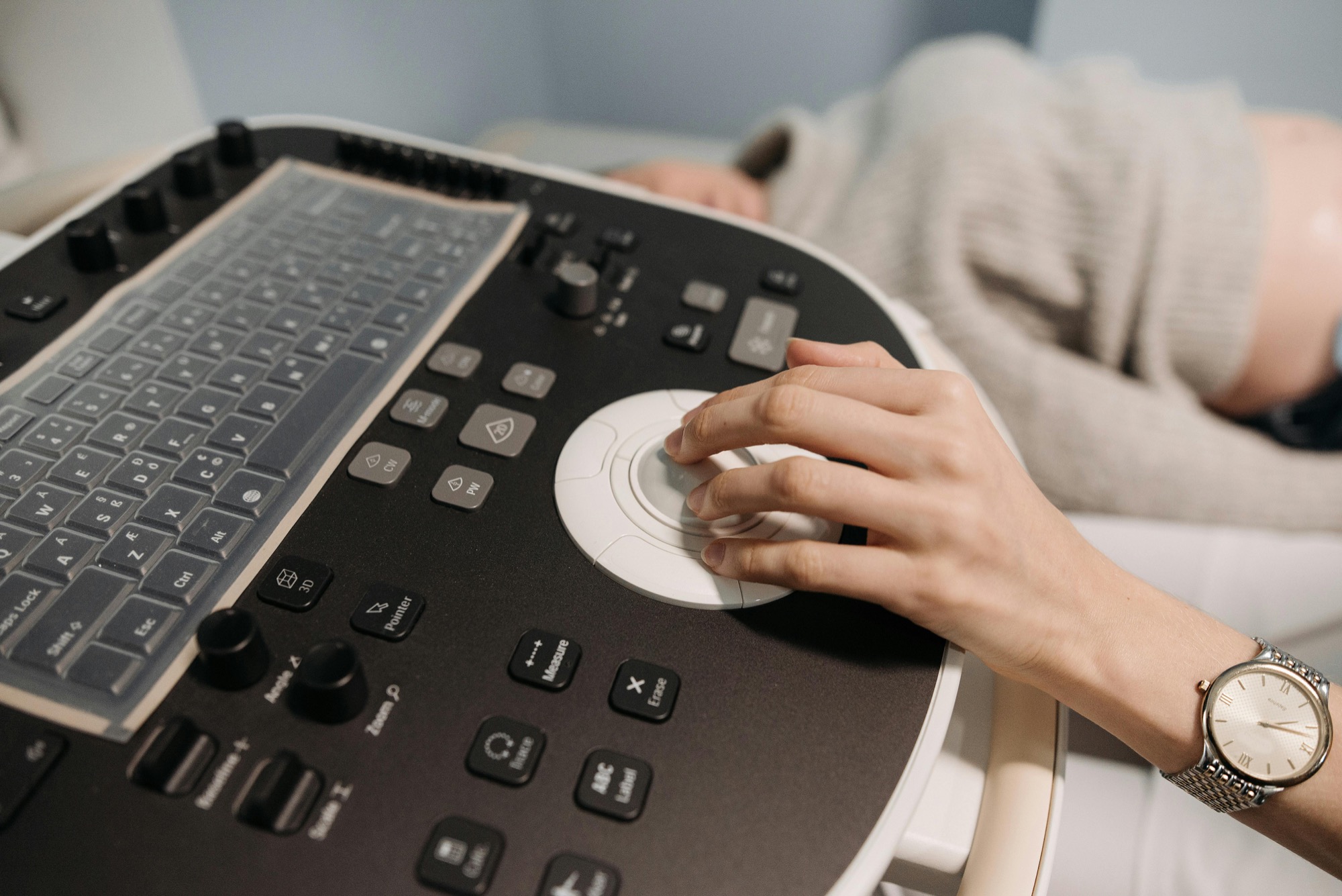
ALARA: The Practical Principle Behind Prudent Use
ALARA means “as low as reasonably achievable.” In ultrasound, it applies to controls that affect acoustic output as well as transducer dwell time and total scanning time. The goal is to obtain the needed image efficiently without using more output or more time than the task requires.
The American Institute of Ultrasound in Medicine’s ALARA statement advises users to begin with appropriate presets, use the lowest output that provides the required image, monitor the thermal and mechanical indices, move or lift the transducer when stationary imaging is unnecessary, and minimize overall scanning time.
For an elective studio, that translates into a prepared operator who knows the machine, scans with a specific purpose, and does not extend exposure simply to chase a difficult view.
Choose the correct preset
Start with a suitable obstetric preset and review manufacturer guidance for the equipment being used.
Use efficient technique
Improve probe position, client position, gain, depth, focus, and rendering controls before increasing output.
Watch the displayed indices
Monitor TI and MI throughout the session and understand how modes and control changes may affect them.
Limit dwell and total time
Keep the probe moving when appropriate and end the scanning portion once the session’s purpose has been met.
Use modes intentionally
Do not use Doppler or other higher-output modes casually. Training should explain when a mode is appropriate and how it affects output.
Document a repeatable policy
Write down the studio’s safety, cleaning, equipment, session, and referral procedures so the team follows one standard.
Understanding Thermal Index and Mechanical Index
TI and MI are on-screen indicators that help a trained operator make prudent decisions. They are not quality scores, and one universal number does not replace training, manufacturer instructions, or examination-specific guidance.
| Displayed index | What it represents | How it supports operator decisions |
|---|---|---|
| Thermal Index (TI) | An estimate related to the potential for tissue temperature rise under defined assumptions. | Observe the appropriate displayed TI, keep output and dwell time conservative, and consider the stage of pregnancy and tissue in the beam. |
| Mechanical Index (MI) | An indicator related to the potential for nonthermal mechanical effects. | Monitor MI as controls and modes change, and use the lowest practical output for the intended image. |
| Scanning time | The total duration of exposure and the time the beam remains over a particular area. | Use efficient technique, avoid unnecessary stationary scanning, and stop when the purpose of the session has been achieved. |
AIUM publishes detailed recommendations that relate displayed TI values to maximum scanning times in clinical contexts. These tables require proper training and should not be reduced to a single blanket time limit for every elective session. Review the AIUM statement on displayed TI and scanning time.
Does 3D, 4D, or HD Rendering Change the Safety Principle?
2D ultrasound displays a sectional image. 3D ultrasound collects data used to create a volume. 4D displays changing 3D information over time, and HD-style rendering applies lighting and surface-rendering techniques to volume data. These presentation differences can change how an image looks, but they do not remove the need for prudent acoustic-output and scanning-time decisions.
A skilled operator first obtains useful 2D information for orientation, then uses appropriate volume-acquisition and rendering controls. Better images often come from probe angle, fetal position, fluid in front of the area of interest, gain, depth, focus, volume angle, and lighting adjustments. Increasing output or continuing indefinitely is not a substitute for technique.
For a focused explanation of elective versus medical purpose, read our diagnostic and non-diagnostic ultrasound comparison.
Safety Includes More Than Acoustic Output
A complete studio policy also covers the condition and cleanliness of the transducer, equipment surfaces, cables, controls, bed, and other areas touched during a session.
AIUM’s 2025 cleaning guidance states that external transducers contacting intact skin require low-level disinfection and should be cleaned after each use. Studios should follow the equipment manufacturer’s instructions for compatible cleaning products and inspect probes, cables, housings, and lenses for damage.
Routine equipment service, documented cleaning procedures, hand hygiene, fresh linens or protective barriers, and orderly gel handling all contribute to a professional client experience.
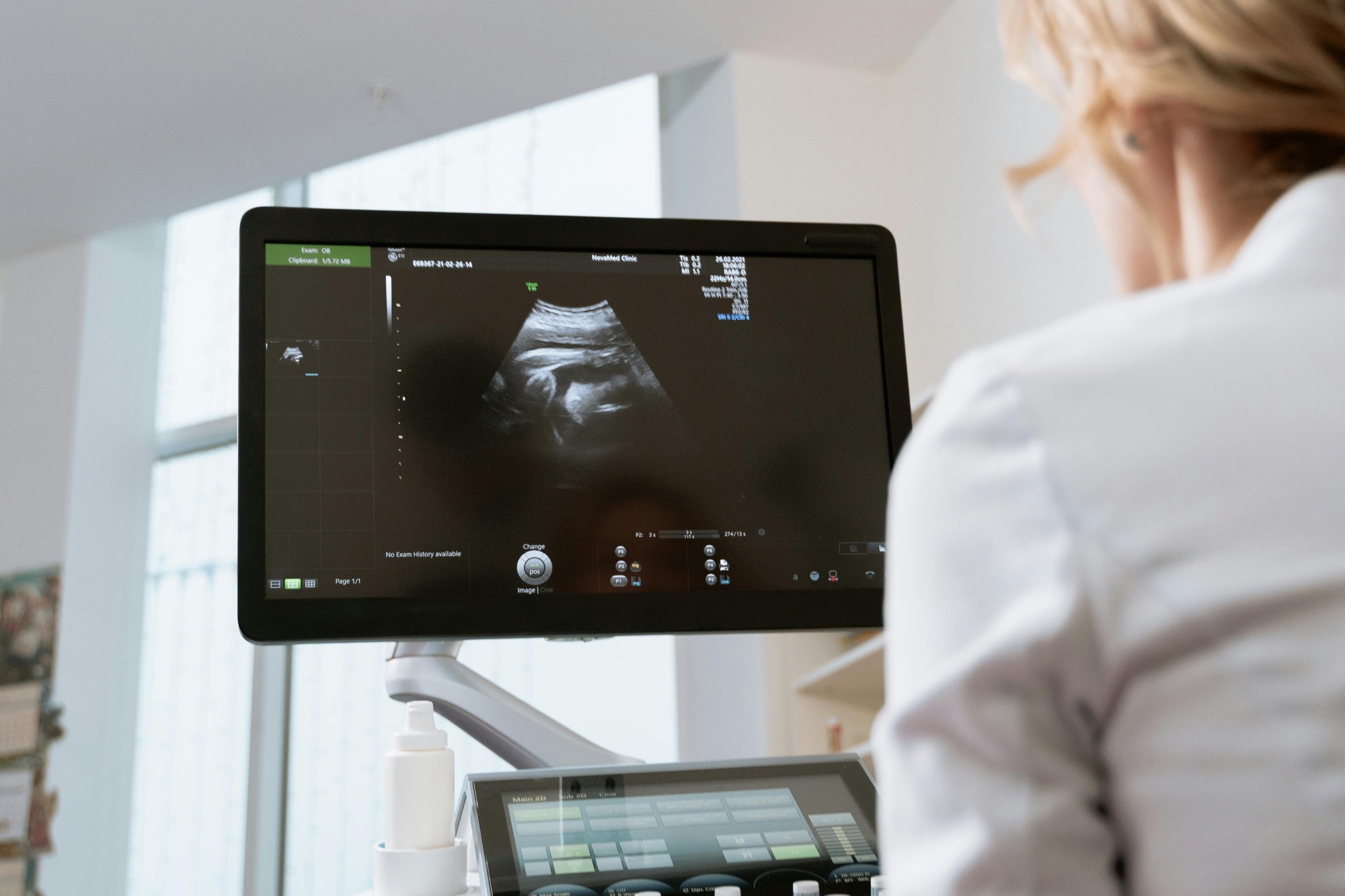
Two Safety Checklists: One for Families and One for Studios
Questions Parents Can Ask
- Is this service clearly described as non-diagnostic?
- What formal ultrasound education and hands-on preparation has the operator completed?
- How does the studio apply ALARA during a session?
- How are the transducer and equipment cleaned between clients?
- How does the studio respond if a client asks a medical question?
- Does the studio encourage continued prenatal care?
- Are session expectations and consent information explained before scanning?
Practices Studios Should Define
- Operator education and supervised practical preparation
- Approved presets, modes, and equipment-use procedures
- TI, MI, dwell-time, and total-time awareness
- Client eligibility, consent, and non-diagnostic disclosures
- A clear pathway for medical questions and unexpected concerns
- Transducer cleaning and equipment inspection records
- Insurance, privacy, maintenance, and incident procedures
Five Common Safety Questions, Answered Carefully
Ultrasound does not use ionizing radiation. It uses sound waves, while still introducing acoustic energy that calls for prudent operation.
Different image presentation does not create a separate safety exemption. 3D, 4D, and HD-style imaging remain subject to appropriate settings, output awareness, and efficient scanning.
There is no responsible universal time promise for every session. Exposure decisions depend on settings, displayed indices, mode, dwell time, and the purpose of the scan.
Modern equipment supports good practice but does not replace operator knowledge. The person scanning must understand the controls, presets, indices, technique, and equipment-care requirements.
Requirements are not identical everywhere. A studio should verify the rules, business requirements, insurance expectations, and service boundaries that apply to its location and operating model.
Elective Imaging Supports, but Does Not Replace, Prenatal Care
An elective session is designed for a non-diagnostic viewing and keepsake experience. It should not evaluate fetal anatomy, growth, placental location, amniotic fluid, pregnancy dating, or another medical concern. Those questions belong with the client’s qualified prenatal healthcare provider.
If a client has pain, bleeding, reduced fetal movement, a concern about pregnancy, or a question about a medical finding, the appropriate next step is to contact their prenatal healthcare provider. An elective studio should not diagnose, interpret, reassure medically, or delay care.
Clear boundaries protect the purpose of the elective experience and help families understand how it fits alongside their established prenatal care.
Frequently Asked Questions
Does ultrasound use radiation?
Ultrasound uses high-frequency sound waves and does not use ionizing radiation. It does introduce acoustic energy, which is why professional guidance emphasizes trained operators and prudent use.
Are 3D and 4D ultrasound different from 2D ultrasound?
They present ultrasound information differently. 2D provides sectional images, 3D creates volume data, and 4D shows changing 3D information over time. The same principles of appropriate output, operator training, and efficient scanning still apply.
What does ALARA mean for an elective session?
It means using the lowest practical acoustic output and the least dwell and total scanning time needed for the session’s purpose. It also includes beginning with appropriate presets and using technique and image controls efficiently.
What are TI and MI?
The Thermal Index and Mechanical Index are displayed indicators that help trained operators consider potential thermal and mechanical effects. They should be monitored in context rather than treated as stand-alone safety guarantees.
Is there one recommended length for every elective ultrasound?
No single time limit accurately covers every combination of mode, setting, displayed index, dwell time, pregnancy stage, and purpose. A trained operator should use conservative settings and keep scanning purposeful and efficient.
How can parents evaluate an elective ultrasound studio?
Ask about the operator’s formal education and hands-on preparation, ALARA procedures, equipment cleaning, non-diagnostic disclosures, prenatal-care policy, and process for directing medical questions to a healthcare provider.
What safety topics should elective ultrasound training cover?
Training should address ultrasound foundations, machine controls, presets, TI and MI, ALARA, dwell and total scanning time, mode selection, probe technique, cleaning, equipment care, non-diagnostic boundaries, consent, and referral procedures.
Build Safety Into Every Part of the Client Experience
Ultrasound Trainers provides private, hands-on elective ultrasound education that connects equipment operation, image acquisition, safety principles, client workflow, and studio preparation.
Review the Training Curriculum Discuss Your Training Goals
How to Get More Bookings for Your Elective Ultrasound Studio in Montana
Struggling to fill your elective ultrasound booking calendar in Montana? This guide covers what actually[...]

Buying an Elective Ultrasound Machine for an Arkansas Studio: What Actually Matters
Ready to buy an elective ultrasound machine for your Arkansas studio? This guide covers the[...]

How to Build a Pre-Launch Marketing Campaign for Your Elective Ultrasound Studio: A 60-Day Countdown Plan
Pre-launch marketing for an elective ultrasound studio begins 60 days before opening. This 60-day countdown[...]

What Career Changers in Hawaii Need to Know About Elective Ultrasound Training
Considering ultrasound training for a career change in Hawaii? This Q&A guide covers what training[...]

How to Find and Recruit Volunteer Scan Models for Elective Ultrasound Practice
Finding volunteer models for elective ultrasound practice is a skill in itself. Learn where to[...]

How to Write an Elective Ultrasound Business Plan That Actually Works
Writing an elective ultrasound business plan? This guide covers what to include, how to build[...]

How to Open an Elective Ultrasound Business in Honolulu: A Practical Startup Guide
Open an elective ultrasound business in Honolulu with confidence. This guide covers training, startup costs,[...]

Adding Elective Ultrasound to an Existing Photography or Doula Practice in Montana
Montana birth photographers and doulas: adding an elective ultrasound add-on business in Montana to your[...]

Starting an Elective Ultrasound Business in Cheyenne, Wyoming: First-Mover Advantage in the Least-Crowded Market in the Country
Thinking about starting an elective ultrasound business in Cheyenne, Wyoming? This guide covers the F.E.[...]
Elective Ultrasound Training for Career Changers in Sweden
Considering a career change into elective ultrasound in Sweden? Find out what training involves, whether[...]

What Career Changers in Fayetteville Should Know About Elective Ultrasound Training
Thinking about elective ultrasound training in Fayetteville, Arkansas as a career changer? This Q&A guide[...]
The Biggest Mistake Las Vegas Elective Ultrasound Studios Make With Their Marketing
Elective ultrasound studio marketing in Las Vegas prioritizes trust and reviews over ad spend. Here's[...]
