

Fixing 4D Ultrasound Artifacts Fast: Shadowing, Speckle & More (2025 Guide)
Why Pristine 4D Images Matter More Than Ever
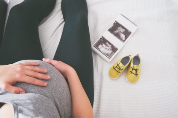
Parents invest in a keepsake baby ultrasound for one main reason: emotion‑packed, jaw‑dropping visuals they can share with family and friends. When shadow bands or snow‑like speckle creep into the live stream, excitement turns to disappointment—and that can ripple through online reviews and repeat bookings. In today’s ultra‑competitive 3D/4D ultrasound business landscape, stellar image quality isn’t a “nice‑to‑have”; it’s a booking engine.
From an operational standpoint, every rescan triggered by poor quality chips away at chair time, increases probe wear, and inflates labor costs. For new studios worried about the cost of starting an ultrasound business, wasted minutes equal lost profit. Even franchise models highlight artifact control as a KPI for profitability, so an independent owner must match or beat that standard to stay ahead.
There’s also the brand angle: crystal‑clear 4D loops serve as the backbone of your social media feed, referral marketing, and website galleries. If your Instagram grid is dotted with noisy frames, you’ll struggle to outrank competitors who’ve mastered their presets. Whether you plan to buy an elective ultrasound machine tomorrow or you’re fine‑tuning a five‑year‑old system, rapid artifact mitigation is a skill set that pays dividends.
Finally, modern families expect an iPhone‑level user experience—instant, intuitive, and flawless. Meeting that expectation starts with equipping every tech on the roster with a quick‑fix playbook they can deploy the second an artifact appears.
The Usual Suspects: Recognizing 4D Ultrasound Artifacts
Before you can fix artifacts, you have to identify them correctly. Below are the five most common image gremlins that plague elective 4D scans. Each description includes visual clues and a short explanation of the underlying physics.
Shadowing
What it looks like: A dark vertical or horizontal band where no anatomy is visible. Bones (especially the fetal skull) are frequent culprits.
Why it happens: Highly attenuating structures absorb or reflect nearly all beam energy, leaving little to return to the transducer. The result is a signal void distal to the object.
Rapid fix: Rotate or rock the probe to find an alternate acoustic window, reduce overall depth to shorten the path length, or activate tissue harmonic imaging. If your console supports multi‑angle compound imaging, toggle it on for challenging heads‑down positions.
Speckle Noise
What it looks like: Grainy snow across the entire ROI that blurs fine detail—especially noticeable on cheeks or tiny fingers.
Why it happens: Interference of returning echoes from multiple scatterers produces a pattern of constructive and destructive signals.
Rapid fix: Lower overall gain, then fine‑tune TGC sliders starting from the top. Engage speckle‑reduction software if available (e.g., GE’s CrossXBeam or Samsung’s ClearVision) and narrow the transmit focus to the structure of interest.
Reverberation (Ring‑Down)
What it looks like: Multiple equally spaced parallel bands beneath a strong reflector—often beneath the maternal abdominal wall or the fetal spine.
Why it happens: Sound waves ping‑pong between two highly reflective interfaces, producing serial echoes at increasing depths.
Rapid fix: Add generous gel to eliminate air gaps, reposition the transducer to alter the angle of incidence, and drop the dynamic range so weaker repeats are suppressed.
Mirror Image Artifact
What it looks like: A duplicated structure appearing deeper than its real counterpart, frequently adjacent to a curved amniotic wall.
Why it happens: The beam reflects off a strong curved interface before striking the anatomy, then bounces back on the same path—making the system miscalculate depth.
Rapid fix: Shift the patient or probe to change angles, use spatial compounding, and reduce depth to confine the potential mirror plane outside the viewing window.
Side‑Lobe Artifact
What it looks like: A ghost echo where no tissue exists—commonly mistaken for an additional limb or mass.
Why it happens: Energy emitted off the main beam axis is reflected back, creating an echo in an incorrect location.
Rapid fix: Increase frequency (narrowing the beam), adjust focus to mid‑ROI, and activate beam‑forming enhancements like multi‑zone transmit.
Your 5‑Minute On‑Console Fix Workflow
Time is money in an elective ultrasound business. The following checklist trains techs to resolve 90 % + of artifact complaints without leaving the exam room:
1 — Check the Fundamentals: Confirm proper transducer selection (4D convex vs. linear), wipe crystal residue off the lens, and add fresh gel. Dirty probes magnify speckle every time.
2 — Reset, Then Rebuild: Hit “2D Default” or your vendor’s baseline obstetric preset. Seasoned techs hate this step—but starting from a known reference eliminates ghost tweaks left by the previous user.
3 — Depth & Focus First: Set depth so baby’s face fills 75 % of the vertical screen, then move the focus arrow just beneath the ROI. This instantly sharpens edges and suppresses side lobes.
4 — Gain, TGC, Dynamic Range: Drop overall gain until the background turns dark gray, then feather TGC to flatten the tissue brightness curve. Finally, narrow dynamic range to ~60 dB for punchy contrast.
5 — Activate Vendor‑Specific Magic: Toggle harmonic or HDLive on GE, TrueVue on Philips, CrystalVue on Samsung. These rendering engines recompute voxel brightness and frequently dissolve residual speckle in real time.
Advanced Troubleshooting & When to Re‑Scan
Some artifacts refuse to die. That’s when deeper settings—and sometimes a full patient reposition—come into play:
Adjust Pulse Repetition Frequency (PRF): Lower PRF reduces aliasing but can exaggerate reverberation. Aim for the sweet spot that shows flow without fuzz.
Modify Persistence: High persistence averages frames and hides random noise but smears motion; drop to low‑medium when scanning active third‑trimester twins.
Zone‑Width Tweaks: Narrowing transmitted beam width boosts lateral resolution, making speckle clusters appear smaller. Most 2024‑2025 consoles expose this control under “Advanced Imaging.”
Patient Repositioning: If posterior placenta or maternal obesity blocks acoustic windows, a side‑lying or hands‑and‑knees position may shift fetal parts into view, erasing shadowing instantly.
Know When to Abort: If BMI 35+ or oligohydramnios severely limits fluid pockets, set clear expectations and offer a complimentary return visit. Forcing a low‑quality capture is a fast track to negative reviews.
Preventive Practices & Ongoing Tech Training
Artifact prevention is cheaper than artifact repair. Schedule quarterly elective ultrasound training refreshers covering new presets, transducer care, and ergonomics. Many ultrasound business training programs bundle image‑optimization drills specific to HDLive, TrueVue, and CrystalVue—don’t skip them.
Build a laminated “Quality First” card attached to each scanning room monitor outlining the five‑step workflow above. New hires absorb it at a glance and veterans appreciate the reminder.
Set up peer‑review huddles every Friday. Techs drop their best and worst loops onto a shared drive, discuss fixes, and keep raising the bar. This simple ritual cuts repeat scans by up to 30 % in many studios.
Finally, audit your console’s firmware. A mid‑cycle update often adds noise‑reduction algorithms at zero cost—an easy win when you’re weighing whether to upgrade a 4D ultrasound machine or stretch its life another year.
Key Takeaways & Next Steps
From instant gain tweaks to advanced beam forming, you now have a toolbox for taming the most notorious 4D artifacts in minutes. Implement the five‑step workflow, keep your transducers pristine, and invest in continuous education to stay ahead of both technological and competitive curves.
Call to Action: Want hands‑on guidance that turns every tech into an image‑quality superstar? Ultrasound Trainers offers on‑site instruction plus virtual refreshers tailored to elective studios. Reach out today and put grainy scans in the rear‑view mirror.
Learn More About Ultrasound Training Learn More About Opening an Ultrasound Studio
What Career Changers in West Virginia Need to Know Before Opening a 4D Ultrasound Studio
Thinking about opening a 4D ultrasound studio in West Virginia as a career changer? This[...]

What Most New Elective Ultrasound Operators Get Wrong Before They Open
The most common mistakes new elective ultrasound studio owners make before opening -- from equipment[...]

How to Start an Elective Ultrasound Business in Minneapolis
Thinking about starting an elective ultrasound business in Minneapolis? This practical guide covers training, equipment,[...]
How Elective Ultrasound Studios in Wichita Build a Consistent Booking Pipeline
Marketing an elective ultrasound studio in Wichita, Kansas? This guide covers local SEO, social strategy,[...]

Elective Ultrasound Training in Hawaii: What You Need to Know Before You Enroll
Considering elective ultrasound training in Hawaii? Learn what programs cover, what the island market demands,[...]

Elective Ultrasound Training and Studio Opportunities in Washington State
Elective ultrasound training in Washington State is within reach. Explore startup opportunities, equipment needs, and[...]

On-Site Elective Ultrasound Training at Your Location: What to Expect and How to Prepare
A complete guide to on-site elective ultrasound training at your location covering curriculum, space preparation[...]

Elective Ultrasound Equipment for Minnesota Startup Studios
Building a keepsake ultrasound studio in Minnesota? This guide covers how to evaluate elective ultrasound[...]

How 3D/4D Ultrasound Technology Has Evolved: From VHS Tapes to HD Live and Beyond
Explore how 3D/4D ultrasound technology evolved from early static 3D volumes through real-time 4D, HD[...]

ARDMS, ARRT, and Non-Diagnostic Credentials: What Elective Ultrasound Studio Owners Actually Need
The ARDMS vs non-diagnostic credential elective ultrasound question confuses aspiring studio owners. This plain-language comparison[...]

Elective Ultrasound Training in France: Regulations, Market Opportunity, and Getting Started
Considering elective ultrasound training France? This guide covers the regulatory landscape, market opportunity in Paris[...]

Networking Strategies for Elective Ultrasound Studio Owners: Associations, Events, and Professional Communities
Networking as an elective ultrasound studio owner builds the peer relationships, referral channels, and operational[...]