

How Elective Ultrasound Works: Technology & Real-Time Imaging Explained
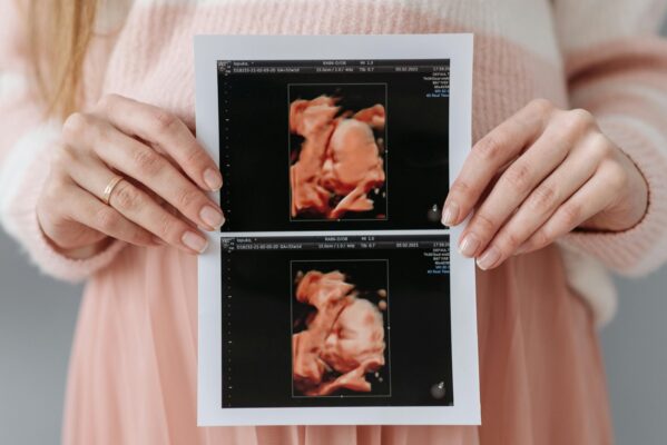
Elective ultrasound is one of the most magical experiences in pregnancy—watching your baby move, smile, blink, stretch, and interact in real time. But beneath the heartwarming images and HD Live renderings lies a fascinating world of physics, advanced computing, and state-of-the-art medical imaging technology.
Whether you’re an expecting parent curious about how 3D/4D/HD ultrasound works or an aspiring business owner learning how to open a 3D/4D ultrasound studio, this deep-dive guide breaks down the science, the equipment, and the imaging process in a way that’s easy to understand.
Let’s explore the exact technology, probes, physics, and rendering engines behind modern elective ultrasound.
What Is Ultrasound? (The Physics Explained Simply)
Ultrasound imaging—whether 2D, 3D, 4D, or HD Live—relies on one core principle:
These echoes are measured by the probe and transformed into real-time images.
Key Terms You Should Know:
- Transducer (Probe): The handheld device that sends and receives sound waves.
- Frequency: The pitch of the sound waves (ultrasound uses extremely high frequencies).
- Echo: The returning sound wave after it bounces off tissue.
- Gain: Adjusts image brightness by amplifying returning echoes.
- Depth: How far the sound waves penetrate into the body.
- Frame Rate: How many images are captured per second.
Unlike X-rays, ultrasound involves:
- No radiation
- No ionization
- No harmful energy
This makes it exceptionally safe for pregnancy imaging.
How 2D Ultrasound Works
2D ultrasound is the foundation of all other ultrasound types.
Here’s what happens during a 2D scan:
- The probe sends sound waves into the abdomen.
- Waves bounce off structures—fluid, soft tissue, bones.
- The machine measures how long echoes take to return.
- A computer converts these measurements into a flat, grayscale image.
2D gives the iconic “side profile” view but doesn’t show lifelike detail. It’s also essential for elective studios because:
- Gender determination begins in 2D
- Baby’s positioning is assessed in 2D
- Technicians use 2D to guide the transition into 3D/4D
How 3D Ultrasound Works: The Science Behind the Still Images
3D ultrasound takes multiple 2D image “slices” and reconstructs them into a 3-dimensional volume.
The process looks like this:
- The volumetric probe scans a wider area than a 2D probe.
- The machine collects hundreds of tiny slices in milliseconds.
- The software stacks and smooths these slices.
- The computer renders the visible surface—usually the baby’s face.
The result is a beautifully detailed, realistic image that parents can study and cherish.
3D Imaging Depends On:
- Baby’s position
- Fluid levels (especially around the face)
- Placenta placement
- Maternal body composition
- Probe angle
- Machine quality
Skilled elective ultrasound technicians learn how to optimize each factor for the clearest images—something taught extensively in training programs like Ultrasound Trainers.
How 4D Ultrasound Works: Real-Time Motion
4D ultrasound is simply 3D ultrasound in motion.
The machine continuously captures 3D volumes and displays them frame-by-frame as video.
4D imaging requires:
- High frame rate
- Fast processors
- High-quality volumetric probes
- Stable fetal position
This is why premium machines like the Samsung HS60 or GE Voluson S8 create buttery-smooth 4D movement while budget machines appear jumpy or laggy.
How HD Live Works: The “Lifelike” Rendering Effect
HD Live (GE) and TrueVue (GE/Samsung) are the most advanced ultrasound rendering technologies available today.
They transform a normal 4D image into a lifelike, photorealistic rendering using digital lighting.
HD Live Adds:
- Virtual light sources (like a spotlight shining on the baby)
- Shadowing that enhances depth
- Soft skin tones generated by algorithms
- Advanced smoothing that reduces artifact or noise
This technology allows parents to see:
- Lips
- Eyelids
- Dimples
- Cheeks
- Expressions
- Fingers and toes
—all with stunning clarity.
The Role of Probes in Elective Ultrasound
Different results depend on different probe designs:
1. 2D Convex Probe
- Used for gender scans
- Used to locate baby’s face
- Used for early heartbeat sessions
2. 3D/4D Volumetric Probe
- Captures volumes—essential for 3D/4D/HD Live
- Has a wider field of view
- Uses specialized crystals to sweep across a large area quickly
These probes alone often cost $6,000–$15,000, which is why quality machines matter.
The Ultrasound Machine: The “Brain” Behind the Images
While probes collect data, ultrasound machines process and render it. Modern machines include:
- AI-based imaging algorithms
- Denoising engines
- Rendering chips for HD Live
- Motion optimization tools
- Dynamic lighting systems
Premium machines used in elective studios include:
- Samsung HERA W10
- Samsung HERA W9
- GE Voluson E8
- GE Voluson E10
- Mindray Z60
- Mindray Z70
How Technicians Get Clear Images: Professional Techniques
Beautiful images don’t happen by accident. They require:
- Correct depth
- Proper angle
- Optimal fluid pocket
- Precise focus adjustment
- Shadow management
- Maternal breathing coaching
This is why professional elective ultrasound training is essential. Without it, image quality will be inconsistent—and sometimes impossible.
Top Technician Tricks Include:
- Having mom lie slightly on her side to move the placenta
- Using gravity to reposition baby
- Having mom drink cold water to encourage movement
- Adjusting frequency for deeper penetration
- Using slow, gentle probe sweeps
These are learned skills—not something a machine does automatically.
Why Some Babies Are Easy to Scan—and Others Aren’t
Baby positioning affects everything.
Good positions:
- Facing outward
- Good fluid pocket around the face
- Head up or sideways
Difficult positions:
- Facing the spine
- Face in placenta
- Face pressed against uterine wall
- Hands or feet blocking the face
Skilled technicians know how to gently work with positioning—not force it.
The Entire Process: Start to Finish
Here’s what happens during a typical elective ultrasound:
- Client checks in and completes non-diagnostic waiver
- Technician reviews baby’s gestational age and session goals
- Warm gel is applied
- 2D scan begins to locate the baby
- Transition into 3D/4D mode
- Adjustments made for clarity, lighting, and angle
- HD Live rendering applied
- Images and videos captured
- Client chooses prints, heartbeat animals, keepsakes
The entire experience is designed for bonding—not diagnosis.
Frequently Asked Questions
No. It uses the same sound waves—only processed differently.
How do you get clear images?
Through positioning, fluid pockets, probe angles, and expert technique.
Do elective machines differ from medical machines?
The technology is the same, but elective studios focus on 3D/4D/HD presets.
Is this technology safe?
Yes—ultrasound uses sound waves, not radiation, and follows ALARA guidelines.
Do technicians need training?
Absolutely. Professional training from programs like Ultrasound Trainers is essential for image quality and safety.
Final Thoughts: The Technology Behind the Magic
Elective ultrasound combines science, physics, software engineering, and human expertise to create breathtaking images that families cherish forever. Modern machines have transformed what’s possible, allowing parents to see their baby with clarity that was unimaginable just a decade ago.
Understanding how ultrasound works gives parents confidence—and empowers future studio owners to operate safely, ethically, and professionally.
Join the Conversation
Did this help you understand how ultrasound works? Drop your questions below! And if this guide inspired you, share it with expecting parents or future elective ultrasound entrepreneurs.

Training Only vs Turnkey Ultrasound Business Package: Which Fits You?
Compare training only vs turnkey ultrasound business package options to see which path fits your[...]

How Sonographers Can Start Their Own Elective Ultrasound Studio
Sonographers have strong technical skills but often misunderstand what owning an elective studio requires. This[...]
Average Revenue of a 4D Ultrasound Business: A Complete Planning Reference
What's the average revenue of a 4D ultrasound business? This complete guide covers revenue variables,[...]

Best Portable 4D Ultrasound Machines for Elective Ultrasound Startups: What to Know
Considering a portable 4D ultrasound machine for your elective startup? This guide separates the myths[...]

Hands-On vs Online Elective Ultrasound Training: Which One Fits You?
Compare hands-on vs online elective ultrasound training for future studio owners. See what each format[...]

Why Your 3D 4D Ultrasound Images Look Blurry and How to Fix It
Struggling with blurry 3D 4D ultrasound images? This step-by-step guide covers the most common causes[...]

Who Can Legally Own an Elective Ultrasound Business? What the Rules Actually Say
The legal ownership question for elective ultrasound businesses is more open than most people assume.[...]

Starting an Elective Ultrasound Business Part-Time: A Realistic Guide
Starting an elective ultrasound business while keeping another income source is a smart approach for[...]

How to Finance an Elective Ultrasound Machine: A Step-by-Step Guide for Studio Owners
Learn exactly how to finance an elective ultrasound machine. This step-by-step guide covers loan types,[...]

Can a Med Spa Add Elective Ultrasound Services? What It Actually Takes
Med spas considering elective ultrasound as an add-on service need to understand training, equipment, compliance,[...]

Ultrasound Business Income Breakdown by Location and Pricing: What Changes Your Monthly Revenue
Ultrasound business income breakdown by location and pricing. See how market, packages, overhead, and positioning[...]

How Profitable Is a Keepsake Ultrasound Business? What Really Drives Profit
How profitable is a keepsake ultrasound business? See what affects revenue, margins, overhead, and long-term[...]