
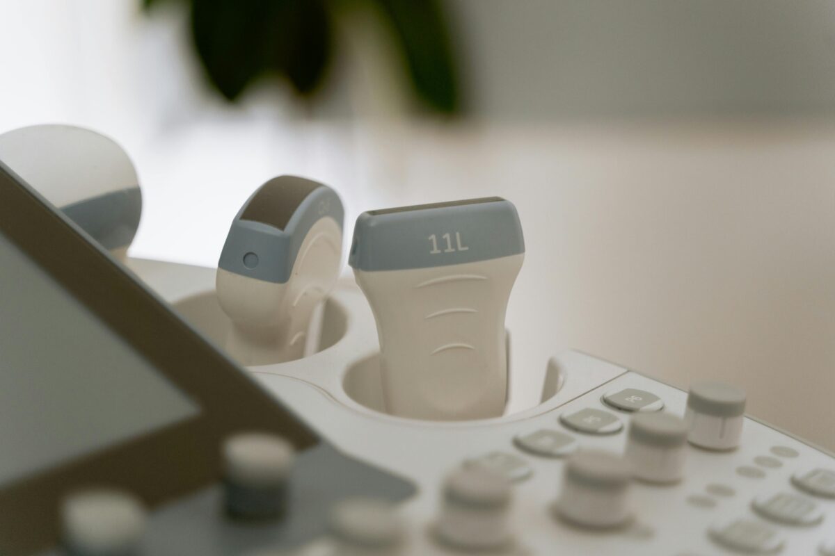
Mastering Probe Positioning: 5 Advanced Techniques for Crystal Clear 3D Scans
Consistent, high quality 3D volumes depend on more than just a premium machine—it’s the art of probe positioning. In this guide, drawn from expert insights at Ultrasound Trainers, we explore five advanced techniques that transform good scans into crystal clear keepsakes. Whether you’re in elective ultrasound training or running a full scale imaging studio, these methods will sharpen your skills and delight clients.
Technique 1: Optimal Probe Angle Alignment
Accurate angle alignment starts with patient positioning. Tilt the probe gently until the target anatomy sits perpendicular to the sound beam. This reduces signal loss and improves volumetric reconstruction.
Use small incremental adjustments rather than large sweeps. A 5° shift can dramatically enhance lateral resolution, making subtle structures pop in 3D renderings.
Train assistants to support the probe at consistent angles during extended sweeps. Stability avoids motion artifacts—critical in elective ultrasound workflows where time is precious.
Document your preferred angle for each anatomical view. Over time, these reference angles become part of your studio’s standard operating procedures, streamlining training and quality control.
Subtechnique: Using Laser Guides for Precision
Laser alignment tools attach to probe housings, projecting a beam that visually indicates your insonation path. This ensures exact repeatability across patients.
Combine laser guides with practice scans on phantoms. By correlating the guide line with actual image orientation, sonographers internalize correct hand positions.
In 3d ultrasound training sessions, integrating laser guidance accelerates learning curves—new operators achieve professional quality images in fewer supervised hours.
Technique 2: Depth and Focus Zone Mastery
Setting your depth just beyond the structure of interest tightens the near field, boosting frame rate and image clarity. Avoid excessive depth that dilutes resolution.
Adjust focus zones to align with the midsection of the target. Multiple focus zones can sharpen layered anatomy but may slow frame rates—use judiciously.
For fetal heart studies, split focus zones at the atrial and ventricular planes. This dual focus method yields crisper volumes for cardiac assessment and keepsake images alike.
During studio training, simulate varied patient sizes. Teach operators to rapidly modify depth and focus settings, ensuring consistency across diverse body habitus.
Subtechnique: Dynamic Depth Adjustment on the Fly
Enable continuous depth mapping when scanning moving targets. This feature automatically tweaks depth settings as you sweep, maintaining image uniformity.
Combine dynamic depth with real time feedback loops: operators watch B mode histograms to confirm optimal penetration and gain before initiating 3D sweeps.
In advanced workshops, Ultrasound Trainers instructors demonstrate dynamic depth on live models, illustrating the immediate impact on volumetric resolution.
Technique 3: Controlled Sweep Patterns for Volumetric Consistency
Standardize your sweep speed: too fast causes data gaps, too slow generates motion blur. Aim for a smooth, even motion of approximately 5–7 seconds per 60° sweep.
Use wrist pivoting rather than arm movement for subtle control. This microadjustment technique reduces fatigue and improves repeatability across long scan sessions.
Mark sweep start and end points on the probe grip with tactile indicators. Operators can then deliver identical sweeps, critical for training protocols and quality audits.
Review recorded volumes side by side during staff meetings. Analyze sweep consistency metrics—angle uniformity, speed variance—to refine team performance.
Subtechnique: Layered Sweep Acquisition
Perform overlapping sweeps that cover the same anatomy at slightly offset angles. This multi angle approach fills in data gaps and smooths surface reconstructions.
Layered sweeps excel in cases with challenging fetal positions, ensuring no area is obscured by bone or amniotic fluid shadows.
Integrate layered sweeps into your 3d ultrasound training curriculum. It teaches resilience against patient movement and yields superior keepsake images.
Technique 4: Fine Tuning Gain, TGC, and Doppler Integration
Balance overall gain to achieve mid grey histograms—too low loses detail, too high flattens contrast. Use histogram tools for objective settings.
Time gain compensation (TGC) sliders manage depth based gain. Smooth, ascending curves prevent near field over amplification and deep field dropout.
Integrate color Doppler overlays selectively to visualize vascularity. This can aid in placental or cord assessments and enhance 3D surface renderings.
In hands on training with Ultrasound Trainers, learners practice toggling Doppler modes mid scan, mastering rapid control changes without breaking sweep flow.
Subtechnique: Automated Gain Mapping Tools
Many modern platforms include automated gain mapping that calculates optimal gain and TGC profiles. Validate these presets against manual adjustments.
Use these automated profiles as baselines, then fine tune for individual patient differences. It accelerates training and reduces operator variability.
Collect feedback during training: track time saved versus image quality improvements, refining when to rely on automation versus manual expertise.
Technique 5: Ergonomic Probe Hold and Body Mechanics
Adopt a handshake grip with the thumb resting lightly on the probe’s top surface. It provides stability and allows microadjustments during sweeps.
Maintain neutral wrist posture: avoid excessive ulnar or radial deviation. Poor ergonomics lead to fatigue, which degrades sweep control over time.
Position your body so the shoulder and elbow absorb movement, not just the wrist. This full limb approach reduces strain during high volume scanning days.
Ultrasound Trainers emphasizes ergonomic best practices in every session, reducing repetitive strain injuries and enhancing long term career sustainability.
Subtechnique: Microbreaks and Stretch Protocols
Implement microbreaks every 15 minutes: simple wrist and shoulder stretches restore blood flow and maintain fine motor control.
Document these breaks in your team’s workflow. Brief stretch routines become part of quality assurance, ensuring every scan benefits from peak operator focus.
Incorporate microbreak training into your 3d ultrasound training programs. It fosters healthy work habits and minimizes downtime due to strain injuries.
Conclusion and Next Steps
Mastery of probe positioning combines physics understanding, precise mechanics, and consistent practice. By integrating these five advanced techniques into your workflow, you’ll achieve crystal clear 3D volumes that impress clients and support diagnostic confidence.
Remember to document your protocols, review performance data, and refresh skills through ongoing 3d ultrasound training with experts like Ultrasound Trainers.
Ready to take your imaging suite to the next level? Share your experiences below or reach out for customized training solutions that elevate every scan.
Which technique will you implement first? Let us know in the comments and tag a colleague who needs to see this!

Training Only vs Turnkey Ultrasound Business Package: Which Fits You?
Compare training only vs turnkey ultrasound business package options to see which path fits your[...]

How Sonographers Can Start Their Own Elective Ultrasound Studio
Sonographers have strong technical skills but often misunderstand what owning an elective studio requires. This[...]
Average Revenue of a 4D Ultrasound Business: A Complete Planning Reference
What's the average revenue of a 4D ultrasound business? This complete guide covers revenue variables,[...]

Best Portable 4D Ultrasound Machines for Elective Ultrasound Startups: What to Know
Considering a portable 4D ultrasound machine for your elective startup? This guide separates the myths[...]

Hands-On vs Online Elective Ultrasound Training: Which One Fits You?
Compare hands-on vs online elective ultrasound training for future studio owners. See what each format[...]

Why Your 3D 4D Ultrasound Images Look Blurry and How to Fix It
Struggling with blurry 3D 4D ultrasound images? This step-by-step guide covers the most common causes[...]
Who Can Legally Own an Elective Ultrasound Business? What the Rules Actually Say
The legal ownership question for elective ultrasound businesses is more open than most people assume.[...]

Starting an Elective Ultrasound Business Part-Time: A Realistic Guide
Starting an elective ultrasound business while keeping another income source is a smart approach for[...]

How to Finance an Elective Ultrasound Machine: A Step-by-Step Guide for Studio Owners
Learn exactly how to finance an elective ultrasound machine. This step-by-step guide covers loan types,[...]

Can a Med Spa Add Elective Ultrasound Services? What It Actually Takes
Med spas considering elective ultrasound as an add-on service need to understand training, equipment, compliance,[...]
Ultrasound Business Income Breakdown by Location and Pricing: What Changes Your Monthly Revenue
Ultrasound business income breakdown by location and pricing. See how market, packages, overhead, and positioning[...]

How Profitable Is a Keepsake Ultrasound Business? What Really Drives Profit
How profitable is a keepsake ultrasound business? See what affects revenue, margins, overhead, and long-term[...]