

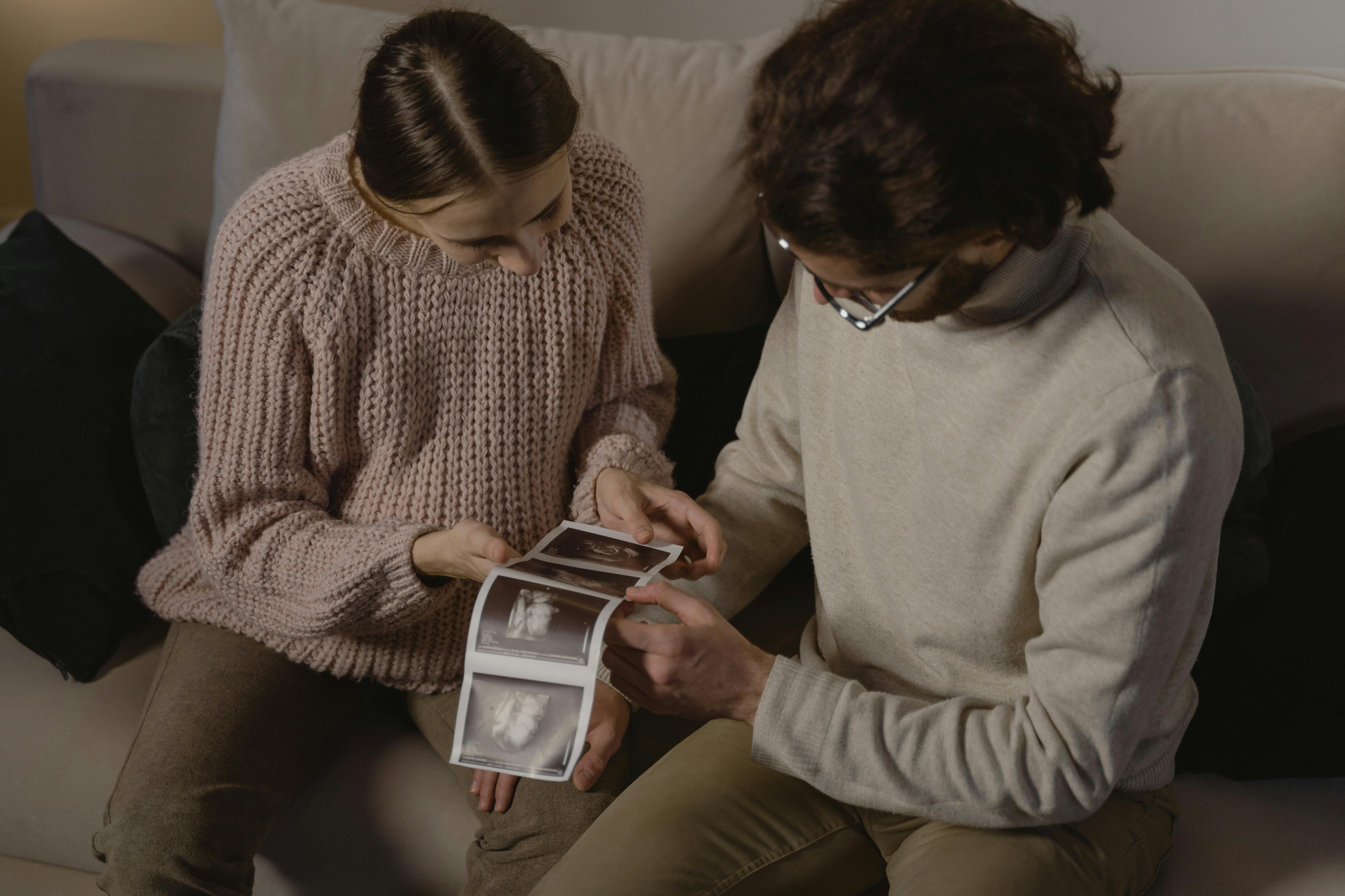
Two technicians can sit behind the same machine, use the same probe, and scan clients of similar gestational age and body type — and produce images that look nothing alike. One session delivers clear, emotionally resonant images that the family will share for years. The other produces something technically functional but forgettable. The machine did not make the difference. The technique did.
Scanning technique in elective ultrasound is not a fixed skill you either have or do not have. It is a craft — one that develops through deliberate practice, attention to what is happening on screen, and a willingness to learn from every session regardless of outcome. The basics can be taught in a few days of quality hands-on training. The refinements take months of consistent practice, genuine curiosity, and honest self-evaluation.
What follows is a collection of the principles and practical insights that experienced elective ultrasound operators build their technique around — the kind of guidance that does not always make it into a training manual because it lives in the accumulated judgment of people who have scanned hundreds of clients across every variable the job can throw at you.
This is not a replacement for formal training. It is the kind of context that makes your training stick — the reasoning behind the technique, not just the steps themselves.
Machine Optimization Is a Skill Unto Itself
Most new scanning technicians underestimate how much is happening before the probe ever touches the client. Your machine’s settings — gain, depth, focus, frequency, and the rendering parameters specific to 3D and 4D modes — are not a set-and-forget configuration. They are active variables that you are adjusting throughout every session in response to what the image is telling you.
A common early pattern is to find a configuration that produces a reasonable image in training and treat it as a default. That default then gets applied uniformly to clients with very different body types, fluid levels, and fetal positions — and the result is inconsistent image quality that reflects the machine rather than the client’s potential. Learning to read your machine’s output and adjust in real time is what closes the gap between acceptable images and excellent ones.
Depth is one of the most frequently misadjusted settings in early-stage scanning. Too shallow and you are missing the context that makes an image readable. Too deep and you are sacrificing resolution in the region that actually matters for a keepsake scan. The right depth for a 28-week scan in a typical client is different from the right depth for a 32-week scan in a client with higher body mass. Adjusting based on what you see — not based on a memorized setting — is the habit to develop early.
Probe Pressure and Angle Are Not Passive Choices
The pressure you apply with the probe, the angle of contact relative to the skin surface, and the tilt and rotation of the transducer are all active variables in every image you capture. Light, even pressure with deliberate angle control tends to outperform the instinct to press harder when images are unclear. Pressing harder does not resolve a positioning or fluid issue — it creates patient discomfort and often degrades the image further by compressing tissue in ways that affect the acoustic window.
Learning to rock and tilt the transducer — rather than just sliding it linearly across the abdomen — is one of the technique refinements that has an outsized impact on image quality. Small angle adjustments change the insonation angle on fetal structures and can dramatically change what appears on screen. This is especially true in 3D and 4D modes, where the depth and angle of acquisition affect how the rendering algorithm builds the surface image.
“The question we are asked most often by operators who are struggling with image quality is whether they need a better machine. Most of the time, the answer is that they need to spend more time learning the machine they already have.”
Reading the Image in Real Time
One of the clearest markers of developing scanning confidence is the shift from watching the probe to watching the screen. Early-stage operators tend to be very conscious of their hand position and movement — which is appropriate in training — but the goal is to reach a point where your primary attention is on what the image is telling you, and your hand is responding to that information automatically.
What the image tells you, if you learn to read it, is a continuous stream of information: where the fetus is positioned relative to the probe footprint, whether you have the acoustic window you need or whether something is obstructing it, whether the depth and gain are showing you what you need to see, and whether a small adjustment in your probe position will give you a meaningfully better result. This is pattern recognition that builds with practice — you cannot shortcut it, but you can accelerate it by being intentional about what you are looking for in every session.
Client Positioning and Preparation Make a Real Difference
The best scanning technique in the world operates within the constraints the client brings to the table. Fetal position, amniotic fluid levels, gestational age, and maternal body composition all affect what is achievable in a given session. But client preparation — what the client does before the appointment and how they are positioned during it — is a variable you can influence.
Adequate hydration in the days before a scan can improve amniotic fluid clarity and give you a better acoustic window. Positioning the client with a slight lateral tilt can shift fetal position enough to improve visibility of the face or the anatomy you are trying to capture. A bolster or pillow under the knees can reduce client discomfort and help them hold a still position longer, which matters in 3D and 4D acquisition where movement creates artifacts.
Communicating clearly with clients about preparation — not just that they should drink water, but specifically how and when — is part of providing a professional service that consistently produces strong results. It is also a way to set realistic expectations: clients who understand that fetal position and fluid levels affect image quality are better prepared to handle sessions where conditions are less than ideal.
Patience in Difficult Sessions Is a Technical Skill
Not every session produces ideal images immediately. Fetal position is one of the most common challenges in elective scanning: the baby’s face is turned toward the placenta, or the position creates a shadow across the features you need to capture, or the baby is simply not cooperating with the view you need. These are not failures — they are normal conditions that every experienced operator encounters regularly.
The technical skill in difficult sessions is knowing what to try in a structured sequence rather than scanning randomly and hoping something changes. Small positional changes for the client, encouraging fetal movement through gentle abdominal stimulation, adjusting acquisition angle, giving the baby time to move without applying pressure — these are deliberate strategies, not luck. Knowing when a session is approaching its productive limit and communicating that honestly to the client is also part of professional technique.
Building a Personal Practice Routine After Formal Training
Formal training is where the foundation is built. The refinement happens in the weeks and months after training through deliberate practice. Scanning on phantoms when live clients are not available keeps your hands familiar with the probe. Reviewing your own saved images critically — looking for what you could have optimized differently — accelerates your visual pattern recognition. Seeking feedback from your training provider or from experienced operators in your network gives you external perspective that self-review alone cannot provide.
The goal of a personal practice routine is not just to stay sharp — it is to keep improving. Operators who treat the end of their formal training as the end of their learning often plateau at a level that produces adequate images. The ones who keep practicing with intention continue improving toward the kind of consistency that produces strong reviews and a reputation for reliable image quality.
FAQ: Elective Ultrasound Scanning Technique
Most operators begin to feel genuinely confident after 20 to 30 real client sessions following their formal training. That timeline shortens with deliberate practice between sessions — scanning on phantoms, reviewing saved images, and actively seeking feedback. It extends if practice between sessions is minimal or passive. The six-month mark is often where operators report a meaningful shift in their sense of ease and consistency.
Pressing too hard with the probe when images are unclear. The instinct when an image is not cooperating is to apply more pressure, but in most cases the issue is not pressure — it is angle, depth, or fetal position. Learning to adjust settings and probe angle before increasing pressure is one of the most important early technique corrections.
Yes. Higher maternal body mass can create a more challenging acoustic window and may require technique adjustments — different probe angles, deeper depth settings, and sometimes a realistic conversation with the client about what is achievable in the session. Professional technique includes honest client communication about variables that affect image quality, not just the scanning itself.
Very important. General scanning principles transfer across machines, but machine-specific optimization — how that machine’s rendering engine responds to different settings, which adjustments produce the most improvement in that machine’s output, and how the software interface is navigated efficiently under session pressure — only comes from time on that specific machine. Training on your actual machine is meaningfully more useful than training on a different model.
You can improve through deliberate self-review and practice, but access to expert feedback accelerates improvement significantly. The most common self-improvement practices are reviewing your saved session images critically, scanning on training phantoms to maintain probe familiarity, and connecting with other operators through training provider networks. Post-training support from your training provider is one of the clearest differentiators between programs that produce confident operators and those that do not.
Serious About Developing Strong Scanning Skills?
Strong technique starts with strong training — hands-on, practical instruction on the machine you will actually use, with support that continues after your training days are complete. Explore elective ultrasound training with Ultrasound Trainers or contact the team to discuss what a training program looks like for your specific situation and goals.
This post was written by the team at Ultrasound Trainers, a company that provides hands-on elective ultrasound training and business support for studio owners across the country. Our training programs emphasize practical scanning technique, machine optimization, and the real-world skills that translate into consistent client satisfaction.

Advanced Scanning Techniques for Elective Ultrasound Operators: Level Up Your Image Quality and Session Efficiency
A detailed guide to advanced scanning techniques for elective ultrasound operators covering gain management, fetal[...]

Elective Ultrasound Studio Interior Design and Client Experience: How Your Space Shapes Your Brand
A practical interior design guide for elective ultrasound studio owners covering the three studio zones,[...]

How to Start an Elective Ultrasound Business in Louisville
Thinking about starting an elective ultrasound business in Louisville? This guide covers training, equipment, location[...]

Elective Ultrasound Training in South Carolina: What You Need to Know
Exploring elective ultrasound training in South Carolina? Learn what the training covers, who it is[...]

Before You Buy an Elective Ultrasound Machine, Read This First
Everything elective ultrasound studio owners need to evaluate before purchasing a machine -- from image[...]

How to Open an Elective Ultrasound Business in Jackson, Mississippi
Thinking about opening an elective ultrasound business in Jackson, Mississippi? This step-by-step guide covers training,[...]

Starting an Elective Ultrasound Business in Wichita: Costs, Steps, and What to Expect
Starting an elective ultrasound business in Wichita takes planning. This guide covers training, costs, and[...]

Starting a 4D Ultrasound Business in Durham, NC: A Real-World Look at the Opportunity
Thinking about starting a 4D ultrasound business in Durham, NC? This story-led guide explores the[...]

Turnkey vs Independent Elective Ultrasound Studio: How to Decide What Is Right for You
Turnkey vs independent elective ultrasound studio compared honestly: what each path includes, what it costs[...]

Hands-On Elective Ultrasound Training Near Kalamazoo and Battle Creek, Michigan
Hands-on elective ultrasound training near Kalamazoo and Battle Creek gives Southwest Michigan birth professionals the[...]

How to Get Your First 10 Ultrasound Clients Fast
Not sure how to fill your booking calendar when you first open? Here is a[...]
Elective Ultrasound Equipment for Tennessee Startup Studios
Building a keepsake ultrasound studio in Tennessee? This guide covers how to evaluate elective ultrasound[...]
Get the Inside Track
Training tips, business advice, and exclusive deals delivered straight to your inbox.
Explore all training formats, durations, and programs in our complete elective ultrasound training guide.